MRI vs. Mammography vs. Ultrasound vs. Blood-Based Testing
Badrinath Konety, M.D.
Chief Medical Officer
Choosing the Right Breast Cancer Screening Tool for the Right Patient
Breast cancer screening has never offered more options or more complexity. For physicians caring for women with dense breasts, elevated risk, or inconclusive imaging findings, determining the next best step often requires balancing diagnostic performance, patient preferences, accessibility, cost, and workflow.
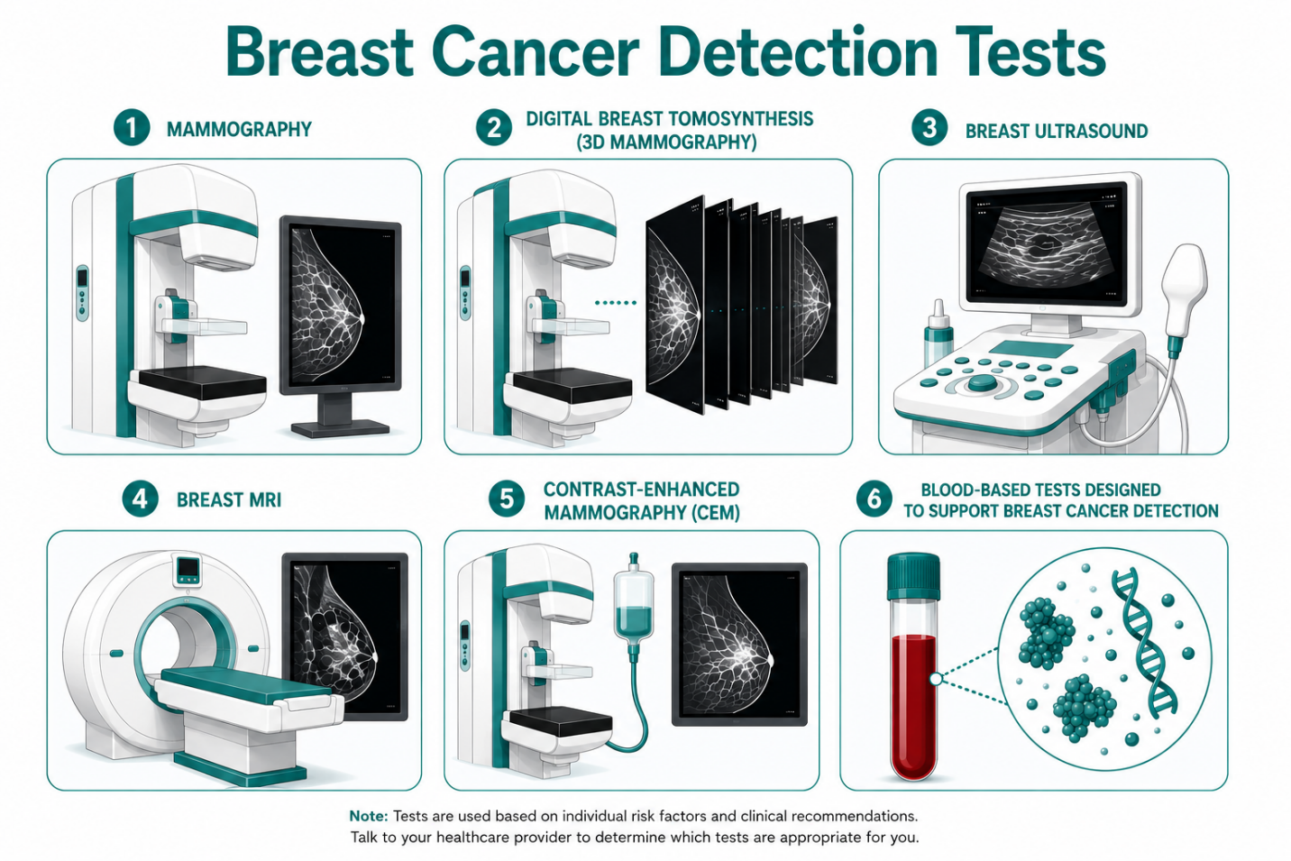
No single screening modality answers every clinical question. Instead, today's approach is increasingly about selecting the right combination of tools based on each patient's risk profile and clinical presentation.
What is a Mammogram? — The Foundation of Breast Cancer Screening
Mammography remains the cornerstone of breast cancer screening and continues to be recommended beginning at age 40 for average-risk women by major organizations including the American College of Radiology (ACR), American College of Obstetricians and Gynecologists (ACOG), and the American Cancer Society (ACS).
Strengths
- Widely available
- Lower cost than advanced imaging
- Proven mortality benefit
- Fast and familiar workflow
Limitations
Dense fibroglandular tissue can obscure cancers, reducing mammographic sensitivity while simultaneously increasing a woman's lifetime breast cancer risk.1 Approximately half of women undergoing screening have heterogeneously or extremely dense breasts.2
For these patients, mammography remains essential but may not always provide the complete picture.
Breast MRI Provides the Highest Sensitivity for High-Risk Patients
Contrast-enhanced breast MRI offers the highest sensitivity among currently available breast imaging modalities and is recommended for many women at elevated lifetime risk.
MRI is particularly valuable for:
- BRCA mutation carriers
- Women with ≥20% lifetime risk
- Certain hereditary cancer syndromes
- High-risk screening programs
Advantages
- Excellent sensitivity
- Superior evaluation of dense breast tissue
- Detects cancers that may be hidden on a mammogram
Considerations
- Higher cost
- Limited availability in many communities
- Longer examination time
- Requires gadolinium contrast
- Higher false-positive rates leading to additional imaging or biopsy
Current guidelines continue to reserve MRI primarily for women meeting high-risk criteria rather than recommending it universally for all women with dense breasts.
How Breast Ultrasounds Are an Accessible Supplemental Option
Whole-breast ultrasound is frequently used in conjunction with mammography, particularly when MRI is unavailable, contraindicated, or dense breast is the only increased breast cancer risk factor.
Benefits include:
- No ionizing radiation
- Greater accessibility than MRI
- Helpful evaluation of focal findings
- Supplemental cancer detection in dense breasts
However, ultrasound is operator dependent and generally has lower specificity than mammography or MRI, resulting in more callbacks and benign biopsies.
For many practices, ultrasound remains an important supplemental imaging tool rather than a replacement for mammography.
Blood-Based Testing: Adding Another Layer of Insight
As precision medicine expands, blood-based diagnostics are emerging as complementary tools that may help clinicians navigate situations where imaging alone leaves uncertainty.
Designed specifically for women with dense breasts, Certitude™ is a blood-based laboratory-developed test intended to support, not replace, mammogram.
Potential clinical scenarios include:
- Dense breasts with a negative mammogram
- Indeterminate or inconclusive imaging
- High-risk screening rotation
- Supporting shared decision-making following abnormal imaging findings
Unlike imaging, a blood draw does not depend on breast density or breast compression and can often be performed during an office visit or at a local laboratory, making scheduling more convenient for many patients.
According to Certitude clinical validation data3:
- >90% sensitivity
- >99% negative predictive value (NPV) in women with dense breasts
A highly reassuring negative result may provide physicians with another evidence-based data point when determining appropriate follow-up, while positive findings should always be interpreted within the context of imaging, clinical evaluation, and established diagnostic pathways.
Rather than replacing mammography, MRI, or ultrasound, blood-based testing represents another layer of clinical insight when standard screening leaves questions.
Clinical Comparison Framework
| Modality | Best Use | Dense Breast Performance | Accessibility | Relative Patient Cost* |
|---|---|---|---|---|
| Mammography | Routine screening | Reduced sensitivity in dense tissue | Excellent | Low |
| MRI | High-risk screening | Excellent | Moderate | High |
| Ultrasound | Supplemental imaging | Operator dependent | Good | Moderate |
| Certitude Blood Test | Supplemental clinical decision support | Not limited by tissue density | Excellent | Moderate |
*Patient costs vary by geography, insurance coverage, and practice.
Selecting the Right Tool
The most effective screening strategy is individualized.
Consider:
- Personal breast cancer risk
- Breast density
- Previous imaging findings
- MRI eligibility
- Patient preferences
- Access to imaging
- Clinical workflow
Rather than viewing these technologies as competing options, clinicians can think of them as complementary components of a comprehensive breast health strategy.
Moving Toward More Confident Care
Dense breast tissue continues to present one of the most common challenges in breast cancer screening. While mammography remains the foundation of screening, supplemental imaging—and increasingly, blood-based diagnostics—may help provide additional confidence when results are less straightforward.
For physicians seeking innovative yet practical ways to support patients with dense breasts, Certitude offers an evidence-based blood test designed to integrate into existing workflows while providing another layer of insight for clinical decision-making.
Learn how your practice can offer Certitude and help provide greater confidence when standard screening leaves questions. Complete our provider inquiry form to learn more.